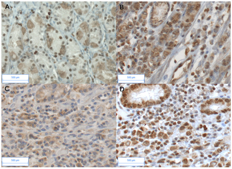
Figure 1.
Immunohistochemical staining of AhR and CYP1B1 in peritumoral and diffuse GCs. AhR in peritumoral gastric tissue (A); weak cytoplasmic and/or nuclear staining were observed in glandular tissue and stroma. In tumoral tissue (B,D), strong AhR immunostaining is observed in most cells, both epithelial and stromal compartments. CYP1B1 (C) and AhR (D) immunostaining are shown on the same tumor (diffuse GC). CYP1B1 was mainly observed in the stromal compartment in diffuse GC (C). Original magnification ×20. Bar scale, 500 μm.

