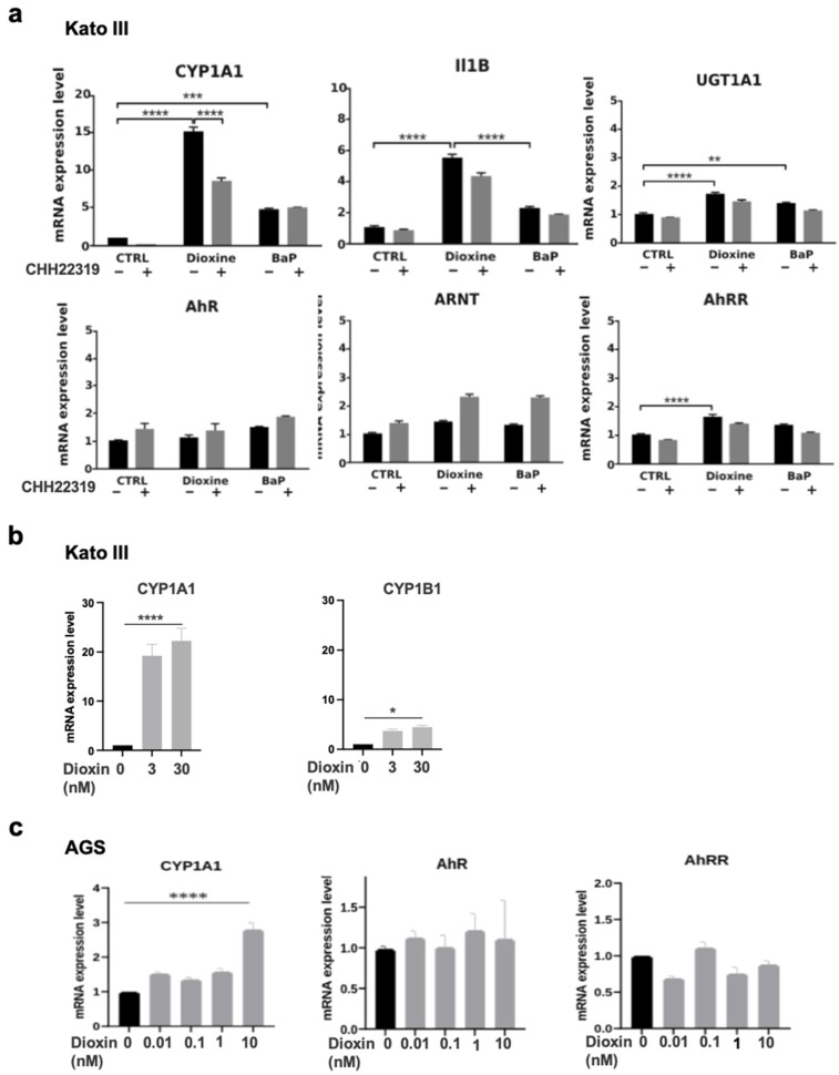
Figure 2.
mRNA expression levels of AhR and AhR-related genes in KATO III and AGS gastric cells upon treatment with either TCDD or BaP. (a) The KATO III cells were cultivated in the absence (Ctrl) or presence of either TCDD (dioxin) 30 nM or BaP (10 µM) for 16 h. The cells were incubated with (gray column) or without (black column) CHH223191 (10 μM). The expression of the indicated genes was determined by qRT-PCR. All the experiments were performed in triplicate. The results are expressed as means +/− S.E.M and normalized so that the mean of the control cells was 1. * p value < 0.005, ** p value < 0.01; *** p value < 0.001; **** p value < 0.0001. (b) The KATO III cells were cultivated in the presence or absence of dioxin at the indicated concentrations. The expression levels of CYP1A1 and CYP1B1 were determined by qRT-PCR in the same experiment. The results were expressed as in (a). (c) The AGS cells were cultivated in the absence (Ctrl in black) or presence of (dioxin) (0.01–10 nM, in gray). The expression levels of the indicated genes were determined by qRT-PCR in the same experiment. All the experiments were performed in triplicate. The results were expressed as in (a).

