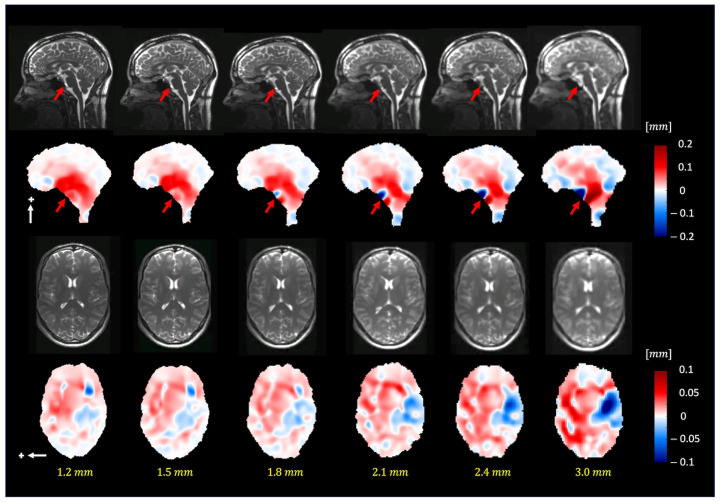
Figure 7.
The pulsatile brain motion in the sagittal (S/I direction, indicated by a white arrow) and axial (L/R direction, indicated by a white arrow) directions for different isotropic spatial resolutions. Plus sign represent the positive direction of motion. As can be seen, the algorithm can robustly estimate the motion field for different image resolutions (up to 1.8 mm isotropic voxel size). Note that the dark blue/red regions (red arrows) in the sagittal plane point to the basilar artery, which exhibits apparent motion (larger than 1.5 pixels).

