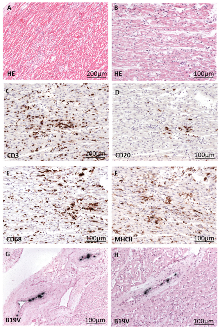
Figure 3.
Histological/immunohistological presentation of fatal myocarditis in a 12-month-old patient (A) with cardiac B19V infection. (A,B) HE staining of heart tissue shows acute myocarditis characterized by myocyte necrosis and extensive inflammatory infiltrate. (C–F) Immunohistochemical staining (brown cells) reveals the presence of many CD3+ T cells (C), some CD20+ B cells (D), and numerous CD68+ macrophages (E), with many of them expressing MHCII (F). (G,H) Detection of B19V DNA (black signals) via radioactive ISH in endothelial cells of cardiac vessels.

