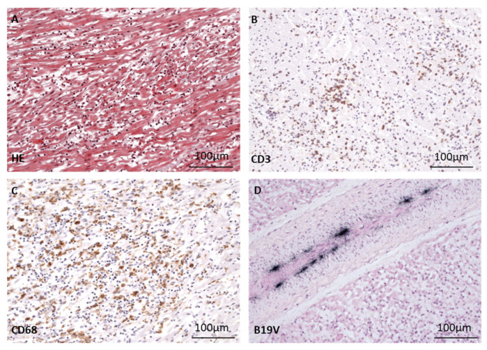
Figure 4.
Morphological presentation of fatal myocarditis in a 17-month-old patient B after cardiac B19V infection. (A) Masson’s trichrome staining of heart tissue shows acute myocarditis with myocyte necrosis, many CD3+ T cells (B) and CD68+ macrophages (C) comparable to findings in patient A. (D) Detection of B19V DNA (black signals) via radioactive ISH in the endothelium of a cardiac vessel.

