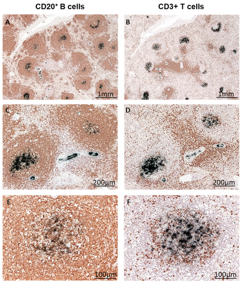
Figure 6.
B19V replication in B cells of the spleen. Splenic tissue from patient B was immunohistochemically stained for CD20+ B lymphocytes (A,C,E) and CD3+ T lymphocytes (B,D,F) (visualized in brown). Consecutive radioactive ISH clearly shows the localization of B19V DNA in B cells (black signal) at different magnifications (A–E).

