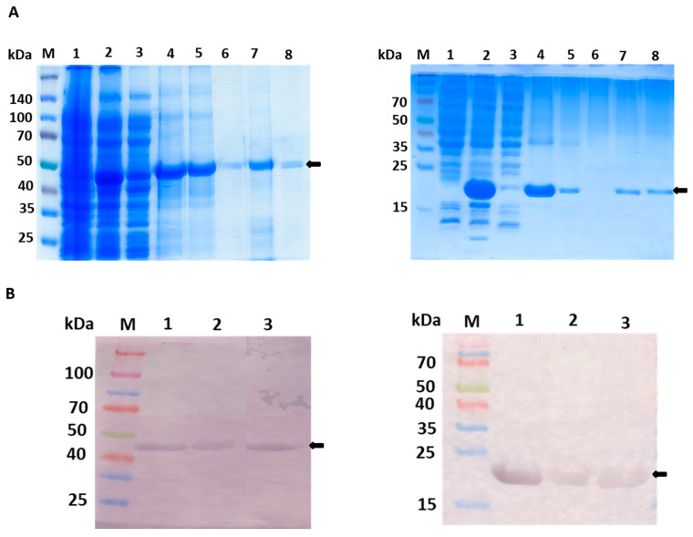
Figure 2.
Purification steps of fGULO-His and cGULO-His using SDS-PAGE (A) and Western blot with anti-His antibody (B). (Aleft) 10% SDS-PAGE of elution fractions from the poly-histidine affinity tag column. M, prestained protein marker with size indicated next to the gel; 1, non-induced lysate of E. coli BL21 (DE3) pRARE transformed with pET28b-fGULO; 2, induced lysate; 3, soluble fraction of lysate; 4, insoluble fraction of lysate; 5, flow through; 6, wash; 7, an aliquot of the pooled eluted fractions; 8, an aliquot of the pooled renatured eluted fractions. (Aright) The 15% SDS-PAGE of elution fractions from the poly-histidine affinity tag column. M, prestained protein marker with size indicated next to the gel; 1, non-induced lysate of E. coli Rosetta (DE3) pLysS transformed with pET28b-cGULO; 2, induced lysate; 3, soluble fraction of lysate; 4, insoluble fraction of lysate; 5, flow through; 6, wash; 7, an aliquot of the pooled eluted fractions; 8, an aliquot of the pooled renatured eluted fractions. (Bleft) M, prestained protein marker; 1, fGULO crude extract; 2, affinity purified fGULO; 3, renatured affinity purified fGULO. (Bright) M, prestained protein marker; 1, cGULO crude extract; 2, affinity purified cGULO; 3, renatured affinity purified cGULO. The arrows indicate the presumed fGULO and cGULO.

