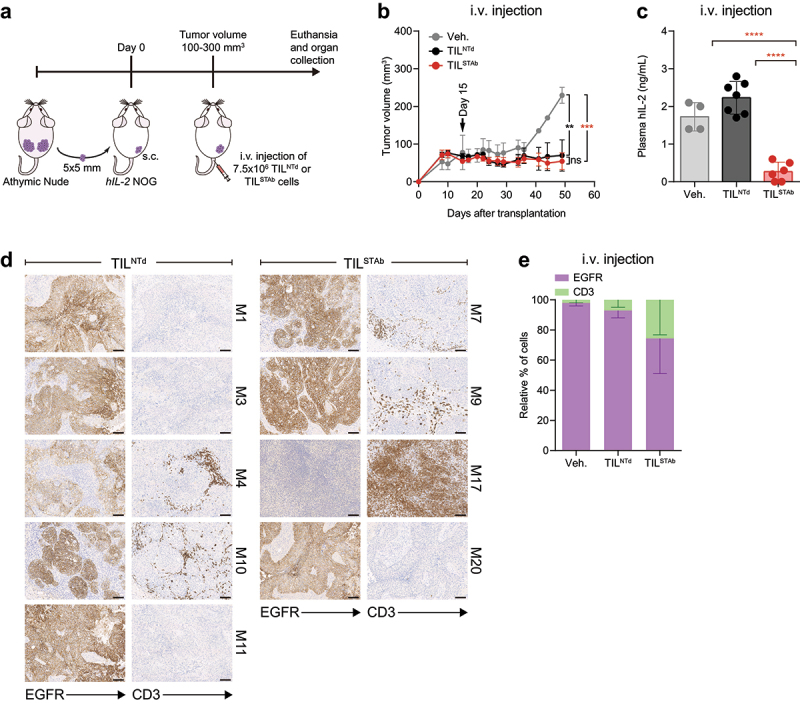
Figure 2.

Treatment of EGFR+ NSCLC PDX mouse model with autologous engineered TCE-secreting TIL. (a) Experimental design. Fifteen hIL-2 NOG mice were xenografted with P1-derived PDX fragments and when the average tumor volume reached approximately 100-300 mm3, were randomized into three groups (2/7/6) and treated i.v. With Veh., (PBS supplemented with 300 IU/mL rhIL-2, n = 2 mice), TILNTd (n = 7 mice) or TILSTAb (n = 6 mice) (7.5 × 10⁶ cells/mouse). (b) Tumor growth curves shown as mean ± SEM; black arrow indicates the day of the i.V. injection. (c) Human IL-2 plasma levels in mice after treatment (mean ± SD). (d, e) EGFR expression and T cell infiltration after TIL treatment by IHC. Scale bars: 100 µm. Data in (e) represent mean ‒ SEM; no significant differences were found. Significance was calculated by two-way (b,e) or one-way (c) ANOVA test corrected by Tukey’s multiple comparisons test. ns, non-significant; **p < 0.01, ***p < 0.001; ****p < 0.0001. PDX, patient-derived xenograft; Veh., vehicle; IHC, immunohistochemistry; SD, standard deviation; SEM, standard error of mean; M, mouse.
