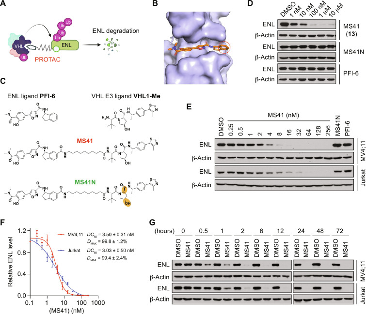
Fig. 1. Discovery of a first-in-class, highly potent VHL-recruiting ENL PROTAC degrader, MS41.
(A) Schematic depiction of VHL-recruiting PROTAC degrader–mediated ENL degradation. Figure was created with BioRender. (B) Cocrystal structure of the AF9 YEATS domain (light blue) in complex with PFI-6 (orange) (PDB code: 8PJ7). The phenyl ring of PFI-6 dihydro indene group (red circle) reaches out to the binding pocket and is solvent exposed. (C) Chemical structures of ENL ligand PFI-6, VHL E3 ligand VHL1-Me, ENL degrader MS41, and MS41N, a negative control compound of MS41. The changed stereocenters are highlighted in orange in MS41N. (D) Immunoblots for ENL in MV4;11 cells treated with DMSO or the indicated concentrations of MS41 (compound 13), MS41N, or PFI-6 for 6 hours. β-Actin was used as a loading control. (E) Concentration-dependent ENL degradation mediated by MS41. Immunoblots for ENL and β-Actin in MV4;11 and Jurkat cells treated with DMSO, the indicated concentrations of MS41, MS41N (256 nM), or PFI-6 (256 nM) for 24 hours. (F) Measurement of DC50 and Dmax values of MS41 in MV4;11 and Jurkat cells. The band intensity in (E) was determined by ImageJ software. Values and error bars are presented as means ± SEM from three independent experiments. (G) Time-dependent ENL degradation mediated by MS41. Immunoblots for ENL in MV4;11 and Jurkat cells treated with DMSO or MS41 (100 nM) for the indicated time. β-Actin was used as a loading control. All immunoblots are representative of three biological repeats.

