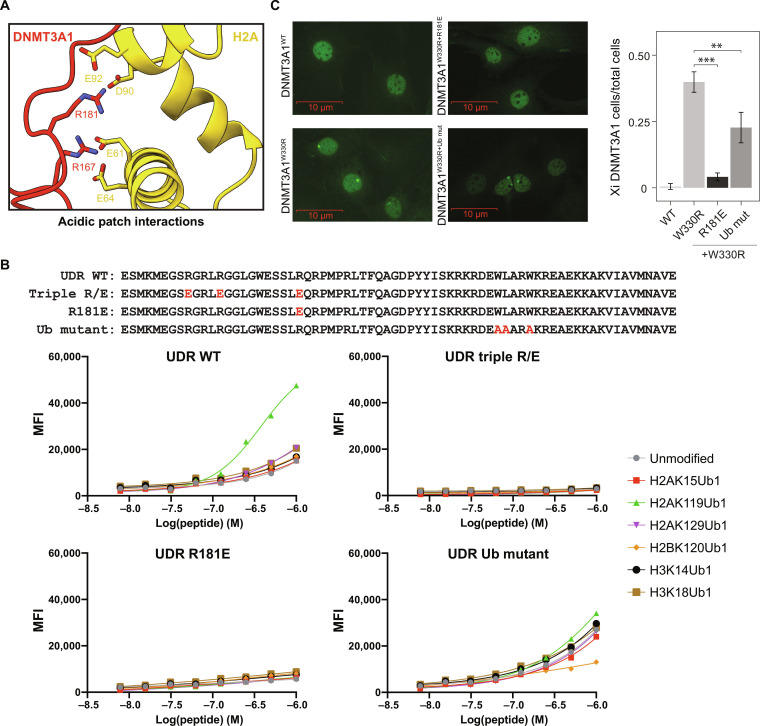
Fig. 2. DNMT3A1 UDR domain binds to H2AK119Ub nucleosome via dual recognition of acidic patch and Ub.
(A) Close-up view of the interactions between the N-terminal region of DNMT3A1 and acidic patch of the nucleosome. (B) Top: Amino acid sequence of the UDR peptide for WT, triple acidic patch mutants (R167E, R171E, and R181E), single acidic patch mutant (R181E), and Ub mutants (W207A, L208A, and W210A). Bottom: dCypher Luminex assay to measure interaction between 6×His-tagged UDR peptides (WT or mutant as noted: the Queries) and a multiplexed panel of fully defined nucleosomes (the Targets; unmodified or with diverse KUb: H2AK15Ub1, H2AK119Ub1, H2AK129Ub1, H2BK120Ub1, H3K14Ub1, or H2BK18Ub1). A four-parameter logistic equation where X is log(peptide concentration) was applied. (C) Left: Representative IF of FLAG-DNMT3A1 in 10T cells expressing DNMT3A1WT, DNMT3A1W330R (PWWP mutant) DNMT3A1W330R+R181E or DNMT3A1W330R+Ub mut. Right: Quantification of FLAG-DNMT3A1 IF in 10T cells expressing DNMT3A1WT, DNMT3A1W330R, DNMT3A1W330R+R181E or DNMT3A1W330R+Ub mut, shown as ratio of 10T cells displaying DNMT3A1 Xi accumulation compared to all 10T cells. Error bars represent SD of five replicates. Student’s t test, **P < 0.01, and ***P < 0.001.

