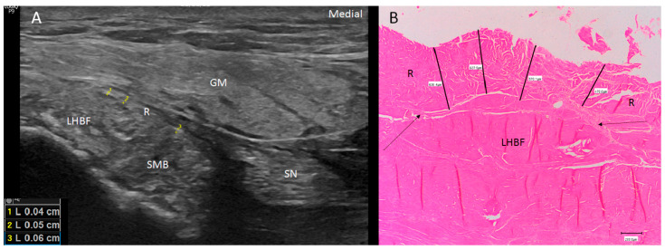
Figure 3.
(A) Ultrasound view of the retinaculum (R) as a hyperechogenic line that fixed them to the bone (as well as the different points where it was measured (yellow numbers 1, 2 and 3). Tendon of long head of biceps femoris (LHBF) and semimembranosus muscle (SMB). Right side: gluteus maximus muscle (GM). (B) Histological view of the retinaculum and its measures of 636.4 µ 637.9 µ, 570.1 µ and 470 µ. It is composed of dense connective tissue, and it is possible to observe the perpendicular direction of its fibers with the fibers of the tendon of LHBF and their close relation (black arrows).

