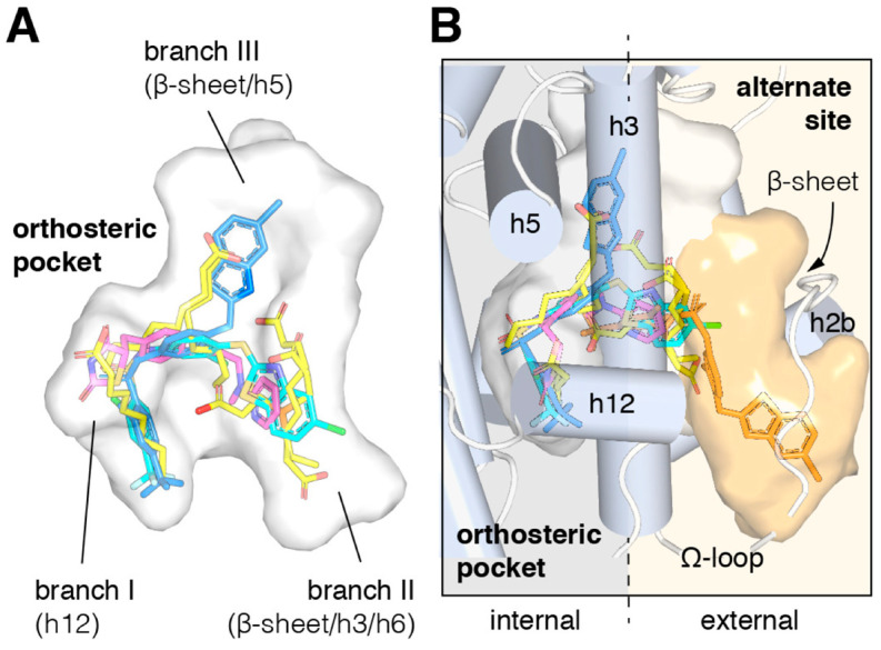
Figure 7.
Structural locations of PPARγ orthosteric pocket and alternate site. (A) The T- or Y-shaped orthosteric pocket can accommodate one or more natural ligands such as nonanoic acid (C9; yellow sticks; PDB: 4EM9) and synthetic ligands such as Rosiglitazone (pink sticks; PDB: 4EMA) or T2384 (light and dark blue sticks representing different crystallized binding modes; PDB: 3K8S, chains A and B, respectively). (B) The orthosteric pocket (the white pocket surface) is completely enclosed within the alpha helical sandwich fold of the ligand binding domain (LBD). Ligands such as T2384 (orange sticks; PDB: 3K8S, chain B) can also bind to a solvent-accessible alternate site (the orange pocket surface) distinct from the orthosteric pocket, structurally defined as the region between helix 3 (H3) and the flexible Ω-loop (the dotted line separating the region internal to the LBD with a gray background and external to the LBD with a light orange background). From “Cooperative cobinding of synthetic and natural ligands to the nuclear receptor PPARγ” by Shang et al. (2018) eLife [81]. This article is distributed under the terms of the Creative Commons Attribution License, which permits unrestricted use and redistribution provided that the original author and source are credited “CC BY 4.0”.

