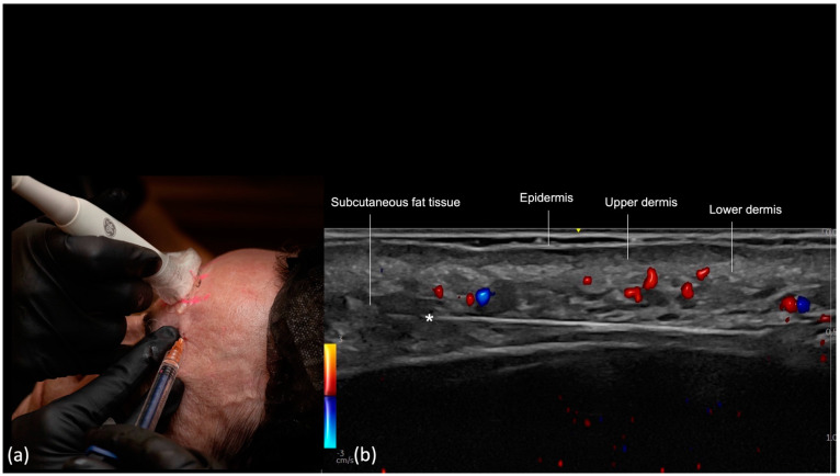
Figure 10.
US−guided injection in the Forehead. The cannula is placed after the region was scanned and the arteries marked (Scan before injecting). (a) Scan while injecting, (b) color Doppler US imaging (LOGIQ E10 GE) is used to ensure the cannula (*) is outside of vessels (in red or blue). Notice the tip of the cannula pointing upwards due to the convexity of the frontal bone.

