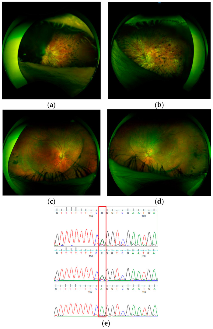
Figure 3.
Ultrawidefield eye fundus images (Optos) - VCAN-related Wagner syndrome family: patient S48, father, 52 years old: (a) (right eye, RE), (b) (left eye, LE); patient S46, son, 20 years old (c) (LE), (d) (RE); (e) Sanger confirmation of the NM_004385.5:c.4004-2A>G variant in VCAN for patient S46 (top), patient S48 (middle), and wild-type control sample (bottom). Due to changes involving the anterior segment of the eye in the father, with extensive phimosis of the lens capsule after cataract surgery, fundus imaging is difficult. In this case, widefield images cover a limited area of the retina; in the right eye, only the nasal part of the retina is visible (a). Pigmentary changes located mainly on the periphery of the retina resemble retinitis pigmentosa, which was the first, initial diagnosis in both father and son. Visual acuity was low at 0.01 (RE), 0.05 (LE), but stable for the last 10 years. In his son, pigmentary and atrophic changes were less severe. The boundary line of the optically empty part of the vitreous body was clearly marked, from which vitreoretinal tractions extend towards the periphery. Best corrected visual acuity is 1.0 (RE) and 0.9 (LE), but with a narrowed field of vision. Snellen charts were used for visual acuity examination.

