
Fig. 3 –
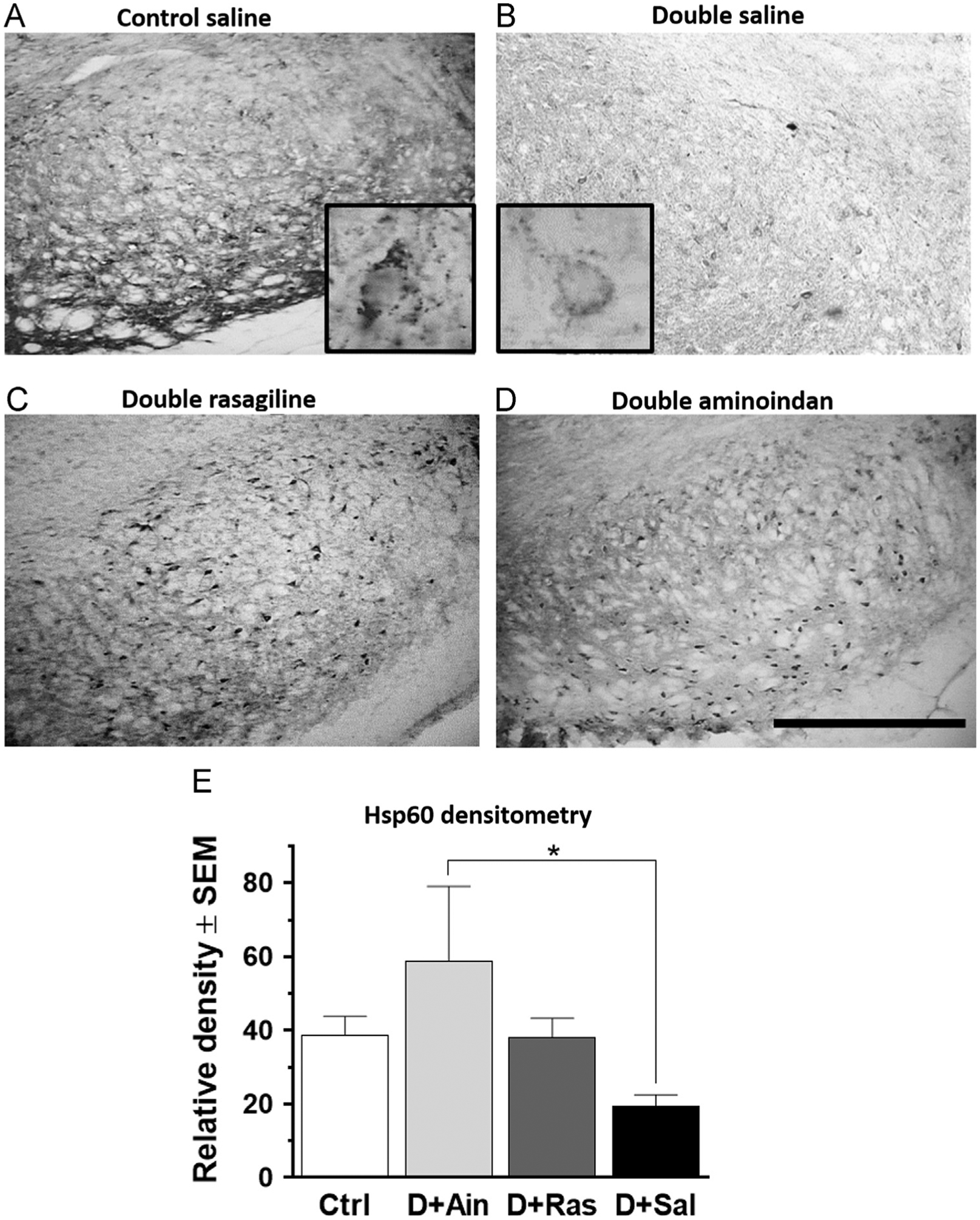
Hsp60 immunostaining in the SN (A–D) and densitometry (E). Hsp60-ir was significantly reduced in the neurons in the SN pars compacta and SN pars reticulata in saline-treated double-lesioned rats (B) compared to control rats (A). Both rasagiline (C) and aminoindan (D) treatments prevented this loss. Insets in (A) and (B) show a higher magnification (60×) of the neuronal morphology for the mitochondrial marker. Bar graph (E) shows densitometry results for the SN and confirms a higher immunoreactivity of Hsp60 in the aminoindan-treated double-lesioned rats (p<0.05) compared to the saline-treated double-lesioned rats. Scale bar in (D) represents 500 μm. Significant differences were tested with a one-way ANOVA followed by Tukey’s post-hoc test (*p<0.05).