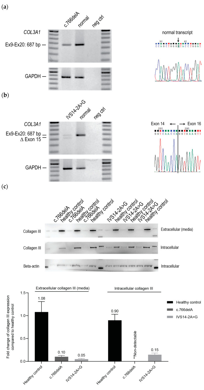
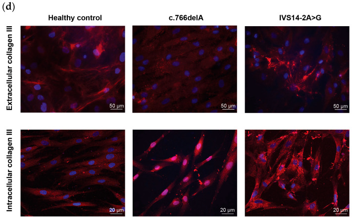
Figure 1.
Characterisation of skin fibroblasts derived from two vEDS patients carrying the mutations c.766delA and IVS14-2A>G. (a,b) RT-PCR amplification and Sanger sequencing of COL3A1 transcripts (exons 9–20) and GAPDH transcript (exon 1–3) from patient fibroblasts. Normal; unaffected healthy control. Neg ctrl; no template control. (c) Western blot analysis of collagen III expression in cell lysate (intracellular) and concentrated supernatant (extracellular). Data from triplicate samples are shown. The bar graph represents densitometric analyses (n = 3, error bars = SD). (d) Immunofluorescence analysis of intracellular and extracellular collagen III in ECM. Red: collagen III. Blue: nuclei.


