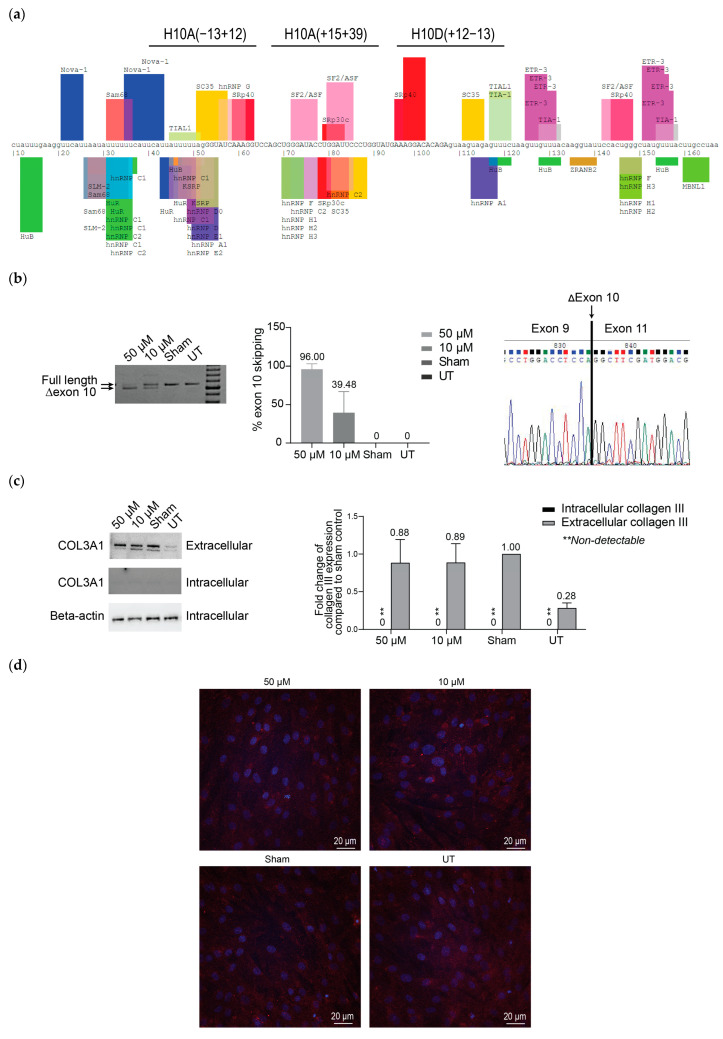
Figure 2.
Assessment of COL3A1 exon 10 skipping and collagen III expression in patient fibroblasts carrying the COL3A1 c.766delA mutation. (a) The exon 10 map indicates the ASO target sites and the exon-splicing enhancer and silencer motifs predicted by Splice Aid. Uppercase letters indicate exonic nucleotides and lowercase letters intronic nucleotides. The image is adapted from the original Splice Aid output (http://www.introni.it/splicing.html, accessed on 19 March 2024). (b) RT-PCR analysis of COL3A1 mRNA across exons 1–14. (c) Western blot analysis of intracellular and secreted collagen III protein. Collagen III is detected at top band; ~140 kDa, however, the presence of lower band is likely a non-specific band due to prolonged image exposure. (d) Immunofluorescence staining of collagen III deposition, (confocal images) in patient fibroblast cultures, transfected with the exon-skipping PMO cocktail [COL3A1_H10A(+15+39) and COL3A1_H10D(+12−13)] at 50 µM and 10 µM for four days. The bar graphs show average values from three independent transfections (n = 3, error bar = SD). Red: collagen III. Blue: nuclei.

