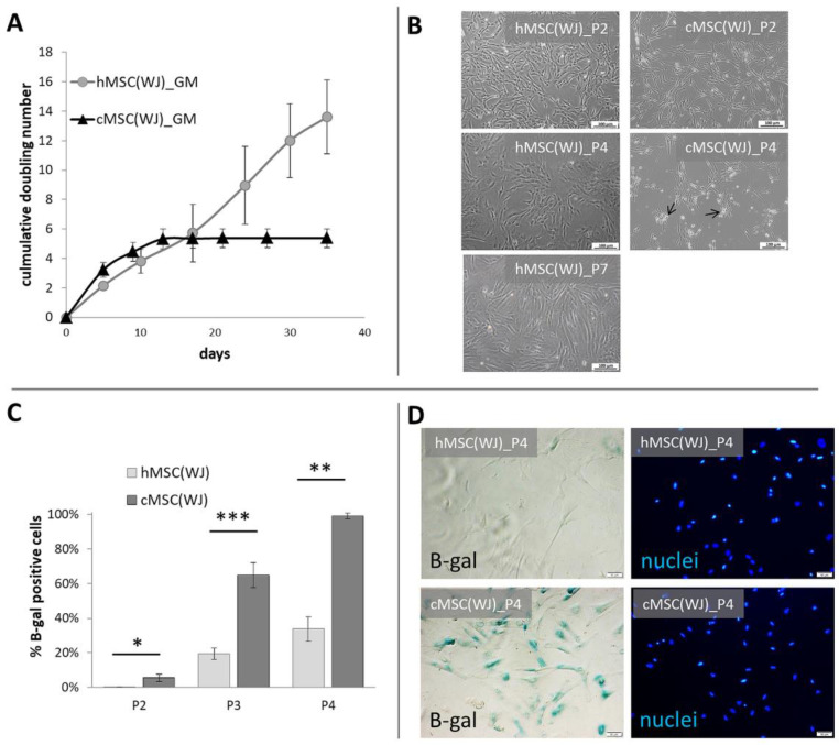
Figure 1.
Growth dynamics and senescence of human and canine umbilical cord-derived MSCs in standard conditions. (A) Cumulative doubling number of representative human (h) and canine (cMSC(WJ)) cells which were cultured for 35 days (day 0—1st passage), (mean values, SD, n = 3). (B) The appearance of human (left column) and canine (right column) cells in standard conditions after the 2nd (P2, upper row),4th (P4, middle row), and 7th (P7, lower picture, human only) passage. Black arrows indicate fragmented cells, scale bars–100 μm (C) The mean (±SEM) proportion of cells displaying senescence associated β-galactosidase (β-gal) activity in human and canine populations on subsequent passages (p). *, p < 0.05; **, p < 0.01; ***, p < 0.001, Student’s t-test, n = 5. (D) Assessment of cell senescence using microscopy: images in rows present the same fields of view. Left column—cells with β-gal activity are blue, right column—cell nuclei stained in blue, scale bars–50 μm.

