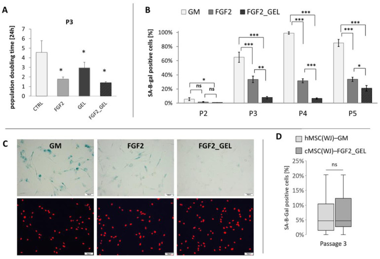
Figure 2.
The effect of different culture conditions on population doubling time (PDT) and senescence of canine Wharton jelly-derived mesenchymal stromal cells, MSC(WJ). (A) PDT of canine MSC(WJ) cultured in growth medium (GM), GM with 2.5 ng/mL FGF2 (FGF2), in GM on gelatin-coated surface (GEL), and GM + FGF2 on gelatin-coated surface (FGF2_GEL), n = 6, *—p < 0.05 in comparison to control using Wilcoxon test or Student’s t-test for related data test depending on data distribution; (B) The proportion of senescent cells within the population (mean ±SEM) cultured in GM, FGF, and FGF2_GEL in subsequent passages (p). Data analyzed within each passage using one-way ANOVA with post hoc Tukey’s test. *, p < 0.05; **, p < 0.01; ***, p < 0.001; n ≥ 5; (C) representative images used for calculation β-gal assay (using X-gal substrate for reaction). Images in columns represent the same field of view. Upper row—light microscopy, senescent cells visible as blue, lower row—fluorescent microscopy, cell nuclei stained in red. Scale bars—50 μm. (D) The comparison of cell senescence at third passage (P3) between human MSC(WJ)s cultured in GM on uncoated surface and canine MSC(WJ) cultured in FGF2_GEL conditions. The graph presents median, quartiles, min and max values, n = 8. Data analyzed using Mann–Whitney U assay. ns—p > 0.05.

