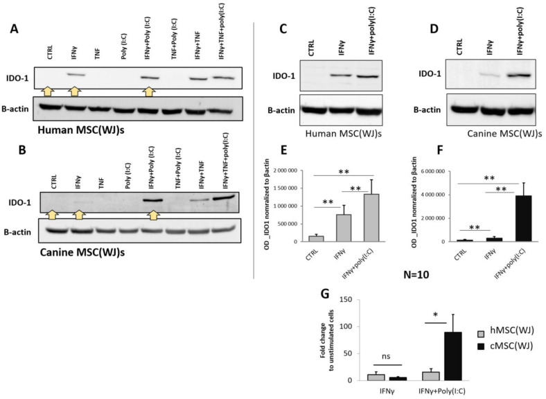
Figure 6.
The effect of pro-inflammatory stimulation on the synthesis of IDO1 in human and canine MSC(WJ)s. Western blot. (A,B) Representative blots presenting IDO1 protein expression in human (A) and canine (B) MSC(WJ)s treated for 24 h with different combinations of IFNγ, TNF, and poly(I:C). Yellow arrows indicate treatments chosen for extended analysis. (C–G) Representative blots presenting the effect of chosen treatments: IFNγ and IFNγ + poly(I:C) in human (C) and canine (D) MSC(WJ)s. (E,F) Mean (±SEM) optical density of IDO-1 normalized to β-actin (n = 10) in hMSC(WJ)s (E) and cMSC(WJ)s (F); 3 independent experiments, cells from 6 different donors in each species, n = 10, analyzed using Wilcoxon test, **, p < 0.01. (G) the comparison of the effect of both treatments on hMSC(WJ)s and cMSC(WJ)s. Mean (±SEM) fold change after treatment in comparison to untreated cells. Mann–Whitney U test, ns—statistically non-significant, *, p < 0.05.

