Figure 6.
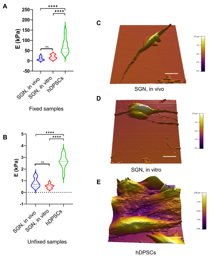
AFM-based nanomechanical characterization of SGN-like cells at 32 DIV. (A) Violin plot of measured Young’s modulus (E) of fixed SGN in vivo, SGN in vitro, and undifferentiated hDPSCs. A significant difference was observed between hDPSCs and SGN samples. However, both SGN in vivo and in vitro were similar related to Young’s modulus measurements. (B) Violin plot of measured Young’s modulus of unfixed SGN in vitro, SGN in vivo, and undifferentiated hDPSCs. A significant difference was also observed between hDPSCs and SGN samples. Young’s modulus measurements of SGN in vivo and in vitro were similar. These measurements indicate strong similarities between SGN in vivo and SGN in vitro differentiated from hDPSCs at the nanomechanical level. (C) Three-dimensional reconstruction of analyzed SGNs in vivo, showing a bipolar morphology. (D) Three-dimensional reconstruction of analyzed SGNs in vitro, showing their bipolar morphology acquired after the in vitro differentiation process, which is different form the morphology of hDPSCs shown in (E). (E) Three-dimensional reconstruction of analyzed hDPSCs showing the characteristic elongated morphology of these cells. Statistical differences were determined with one-way ANOVA. p values are indicated with **** p ≤ 0.0001. n = 10 measurements. Scale bar = 20 µm.

