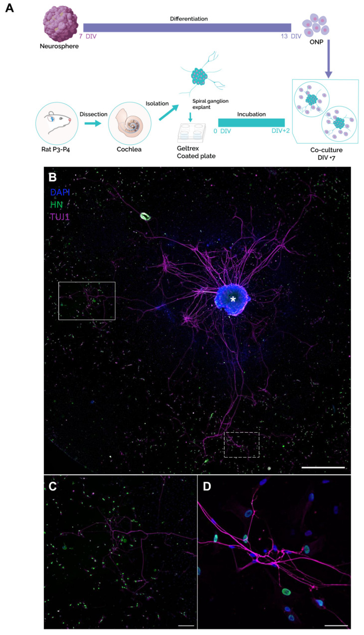
Figure 7.
Characterization of the co-cultures between human ONP cells and rat SG explants. (A) Schematic representation of different steps of the co-culture procedure. Neurospheres were generated from hDPSCs and differentiated to ONP cells until 13 DIV. In parallel, cochlear explants were dissected from the inner ear of postnatal day P3 rats, followed by SG isolation and culture on Geltrex-coated plates for 48 h. Then, the ONP cells were detached from the substrate and co-cultured with SG explants for 5 additional days. (B) Representative image of neurite outgrowths from SG explant (asterisk) immunostained with anti-TUJ1 (shown in magenta), projected towards the ONP cells, immunostained with anti-human nuclei (shown in green). DAPI was used to counterstain the nuclei. Scale bar = 1000 µm. (C) Magnification of the area indicated by the white rectangle in (B, showing outgrowth neurites (magenta) emanating from the SG explant towards ONP cells (green), Scale bar = 500 µm. (D) Magnification of the area indicated by the dashed rectangle in (B) highlights contacts between neurites from SG explant and the membrane of ONP cells. Scale bar = 50 µm. Abbreviations: ONP: otic neuronal progenitors; SG: spiral ganglion.

