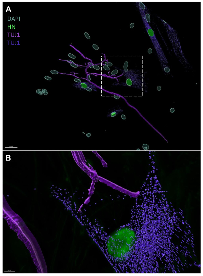
Figure 8.
Three-dimensional reconstruction of SG explant-emanating neurites and ONP contacts using IMARIS. Figure S8B was used for the reconstruction with two look-up tables for TUJ1 staining. (A) Image represents the 3D reconstruction of neurite contacts between SGNs from the SG explant and ONP cells using IMARIS 10 software. The neurites (magenta = TUJ1 immunostaining) establish direct contacts with the membrane of ONP derived from hDPSCs (green: human nuclei, and purple: TUJ1). Scale bar = 30 µm. (B) Magnification of the area indicated by the square in (A) representing the image reconstruction by IMARIS of the neurites which established contacts during the co-culture. Scale bar = 10 µm.

