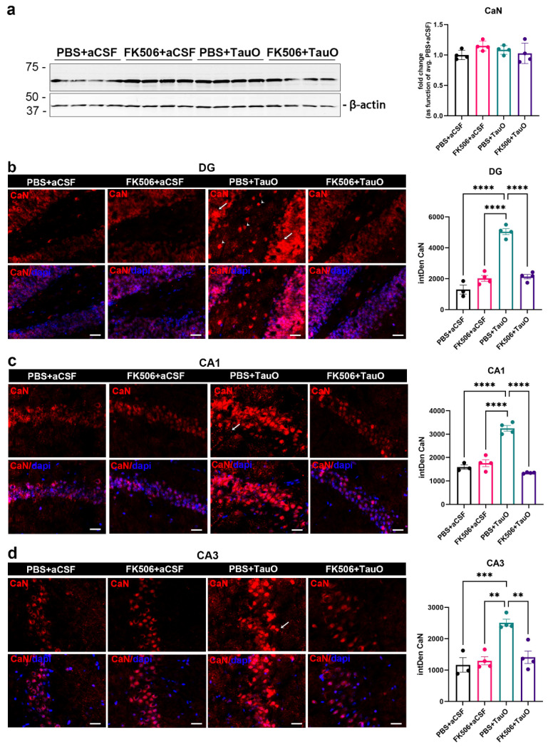
Figure 2.
Effects of FK506 administration on TauO-induced increased levels/visibility of CaN in the mouse hippocampus. (a) Western blot analysis of CaN protein levels across the groups and relative quantification as function of average of PBS + aCSF mice; (b–d) (Left panels) Representative immunofluorescence images of CaN (red) levels in (b) dentate gyrus (DG), (c) CA1, and (d) CA3, from control mice (PBS + aCSF and FK506 + aCSF), mice ICV injected with TauO (PBS + TauO), and mice ICV injected with TauO and treated with FK506 (FK506 + TauO)). Original magnification (60×), scale bar (30 µm) and relative quantitative analyses (right) of the fluorescence intensity across all experimental groups. White arrows and white arrowheads indicate CaN distribution across the hippocampal regions analyzed. (d) Data represent the mean ± SEM; biological replicates n = 4 per group; one-way ANOVA with Tukey’s post hoc corrections for multiple comparison; ** p < 0.01, *** p < 0.001, and **** p < 0.0001.

