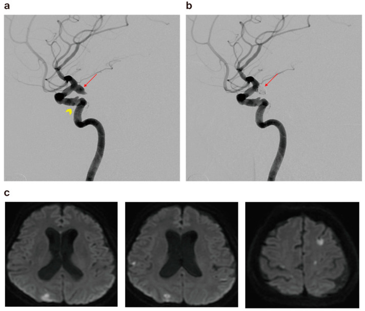
Figure 2.
(a) A 59-year-old female patient’s cerebral angiography revealed ruptured right posterior communicating artery aneurysm (arrow) and atherosclerotic change in the left cavernous ICA (arrowhead). (b) Cerebral angiogram showing minimal residual filling of the neck after coiling (arrow) via the double microcatheter technique. (c) Diffusion-weighted imaging obtained 1 day post embolization, revealing multiple tiny acute infarctions.

