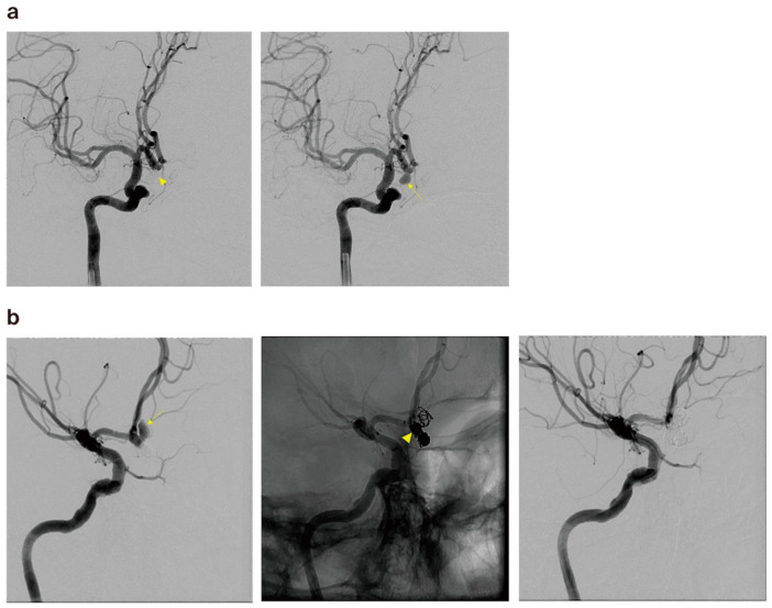
Figure 3.
(a) The cerebral angiography of a 57-year-old male patient revealed a ruptured right anterior communicating artery aneurysm (arrow) and postoperative angiography after coil embolization (arrowhead). (b) The patient presented to the emergency room with SAH at 30 months after the treatment, and cerebral angiography revealed coil compaction and aneurysm regrowth with a newly observed pseudoaneurysm (arrow). Partial coiling was performed on the pseudosac, and compact embolization was performed on the common neck portion (arrowhead).

