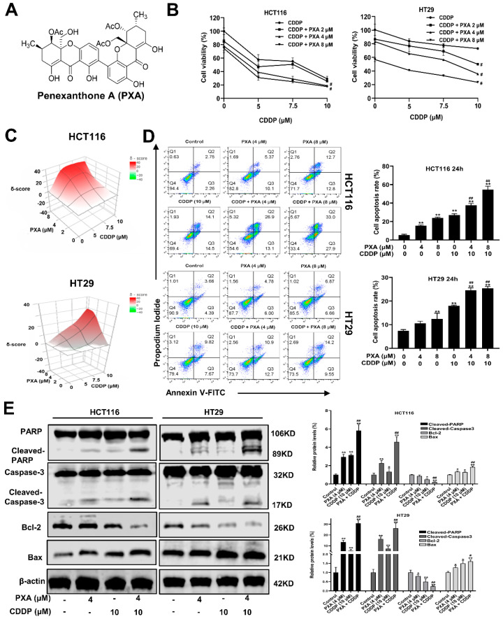
Figure 1.
PXA sensitizes CRC cells to CDDP-induced cytotoxicity and apoptosis. (A) Chemical structural of PXA. (B) CRC cells were co-treated with PXA and CDDP for 48 h, and the percentage of cell viability was determined by CCK-8 assay. (C) 3D visualization of synergy scores between PXA and CDDP obtained using the SynergyFinder tool; these calculated average synergy scores are 20.7 and 11.3 for these two panels of drug combinations (Synergy scores > 10 are considered synergistic). (D) The percentage of apoptotic cells was analyzed and quantified using flow cytometry after Annexin V-FITC/PI staining. (E) The protein levels of Cleaved-PARP, Cleaved-caspase-3, BAX, and Bcl-2 in HCT116 and HT29 cells were detected by Western blot after 24 h treatment; β-acting was used as a loading control. Results are expressed as means ± SD. * p < 0.05, ** p < 0.01 versus the control group, # p < 0.05, ## p < 0.01 versus the CDDP-treatment group.

