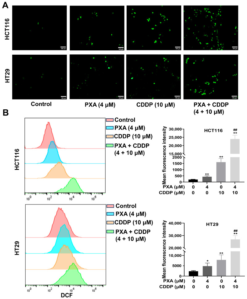
Figure 2.
PXA increased CDDP-induced ROS production. (A,B) HCT116 and HT29 cells treated with PXA and CDDP were incubated with the DCFH-DA probe for 20 min, and the ROS levels (DCF fluorescence) were observed and analyzed by fluorescence microscopy (A) and flow cytometry (B). Scale bars: 100 μm. Results are expressed as means ± SD. * p < 0.05, ** p < 0.01 versus the control group, ## p < 0.01 versus the CDDP-treatment group.

