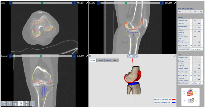
Figure 2.
Three-dimensional planning: The software enables us to conduct three-dimensional planning based on a CT scan. To preserve the native LDFA, we planned a distal femoral cut at 2.5° valgus with a bone thickness of 6 mm. The posterior femoral cut was set at 0° of rotation with a bone thickness of 5 mm. The tibial cut was planned with a varus of 3.5° and a PTS of 8.5°. Additionally, we determined the optimal component sizes, selecting a femoral component size 3+ and a tibial component size 3. In red is represented the position of the planned femoral component, while in blue is the position of the tibial component.

