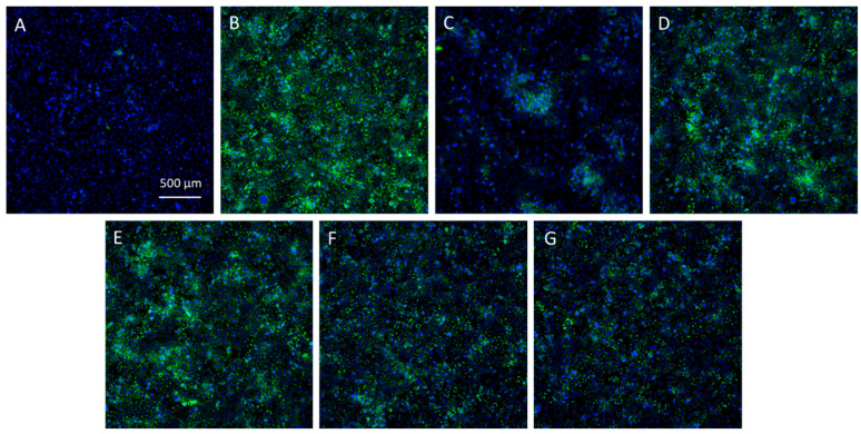
Figure 9.
Microphotographs of SEBO662AR human sebocytes. Intracellular lipid droplets were labelled with the fluorescent probe BODIPY® 493/503 before fluorescence imaging (magnification ×20). The cells were either unstimulated (A) or stimulated with a lipogenic mix containing Dihydrotestosterone, Vitamin D3, Vitamin C, insulin, and Ca2+ ions (confidential concentrations, QIMA Life Sciences lipogenesis inhibition assay) (B–G), followed by treatment with control medium (B), dutasteride at 10 µM (C), cerulenin at 10 µM (D), or the Skm ethanol extract at concentrations of 1 (E), 5 (F), or 10 (G) µg·mL−1. Cell nuclei were stained with Hoechst (blue labeling), while green labeling indicates lipogenesis detected using the BODIPY® probe.

