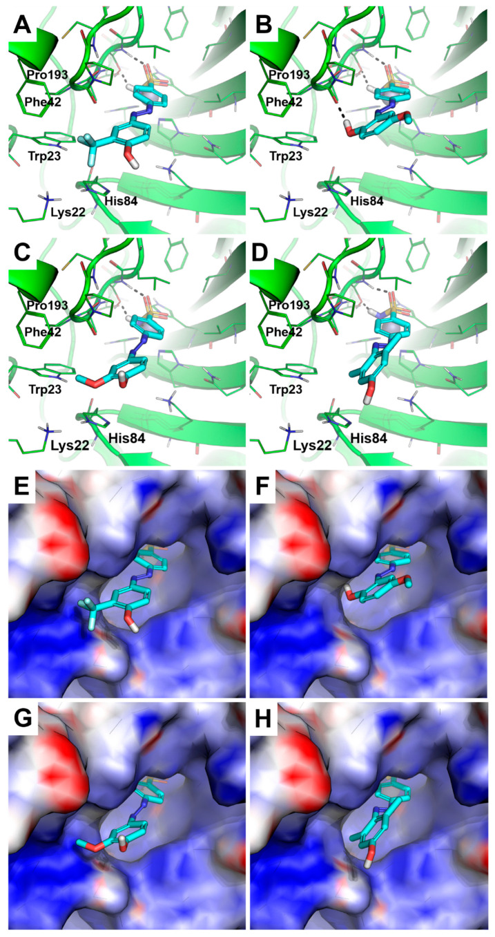
Figure 2.
Predicted binding mode of compounds 4d (A), 4a (B), 4b (C), and 4g (D). The crystallographic structure of HpαCA, coded by PDB ID 4XFW, is shown as the green cartoon and lines in panels (A–D), while the protein surface is colored according to the electrostatic surface potential in panels (E–H) (blue = positive charge, red = negative charge, white = uncharged lipophilic regions). Residues within 4 Å from the ligands are shown as green lines. Those of 4d,a,b,g are shown as cyan sticks; non-polar hydrogen atoms are omitted. The catalytic Zn(II) ion is shown as a gray sphere. Polar interactions between the inhibitors and HpαCA are highlighted by black dashed lines.

