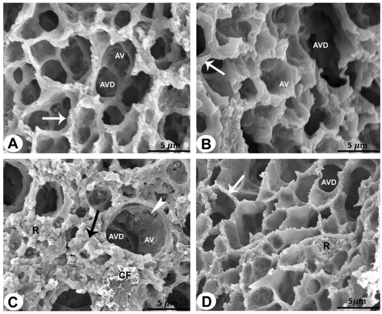
Figure 2.
SEM of the rat lungs of all groups (bar = 5 µm). (A,B) Rat lungs of the control and sham-operated groups showing alveoli (AV) and alveolar ducts (AVD) surrounded by an enhanced alveolar ridge (arrows). (C) Rat lung of the AMD-treated group showing damaged and degenerated alveoli (AV) and alveolar ducts (AVD) of the lung sections. Destruction of alveolar wall lining (arrowheads) and increased amounts of erythrocytes (R) and collagen fibrils (CF) can be seen in the interalveolar septum (black arrow). (D) Rat lung of the LC plus AMD-treated group showing an improvement in the lung architecture. The alveoli (AV) and alveolar ducts (AVD) are surrounded by an enhanced alveolar ridge (arrows).

