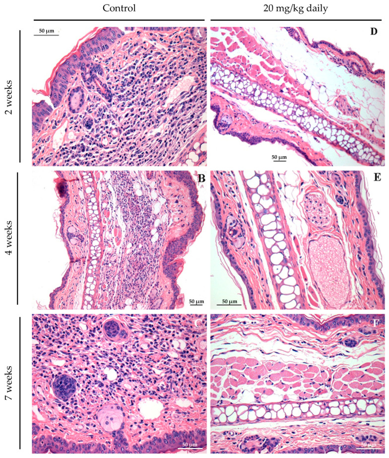
Figure 8.
Long-term assessment of the inflammatory process in the ears of mice infected with L. braziliensis and treated intraperitoneally with 17-DMAG at later stages of treatment. Images represent inflammatory infiltrates in the lesion of BALB/c mice of the kinetic experiment described in Figure 7. After two (A,D), four (B,E), or seven weeks (C,F) after treatment. Control mice received 5% glucose solution intraperitoneally during the same time points. All animals were euthanized, and their ears were removed, embedded in paraffin, cut on a microtome, and stained with H&E for histopathological analysis. Histological sections were analyzed, and the images were obtained under observation in an optical microscope. Magnification 40×.

