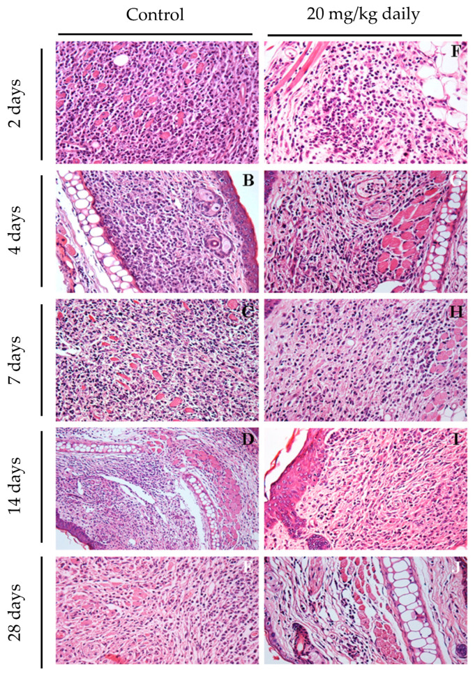
Figure 10.
Early assessment of inflammatory process in ears of mice infected with L. braziliensis and treated with 17-DMAG intraperitoneally. Images represent inflammatory infiltrates in the lesion of BALB/c mice infected and treated as explained above. After 2 (F), 4 (G), 7 (H), 14 (I), and 28 days (J) of treatment. Control mice received 5% glucose solution intraperitoneally during the same time points (2 days: (A); 4 days: (B); 7 days: (C); 14 days: (D); 28 days: (E)). All the animals’ ears were removed, embedded in paraffin, cut on a microtome, and stained with H&E for histopathological analysis. The histological sections were analyzed, and the images were obtained under observation in an optical microscope. Magnification 400×.

