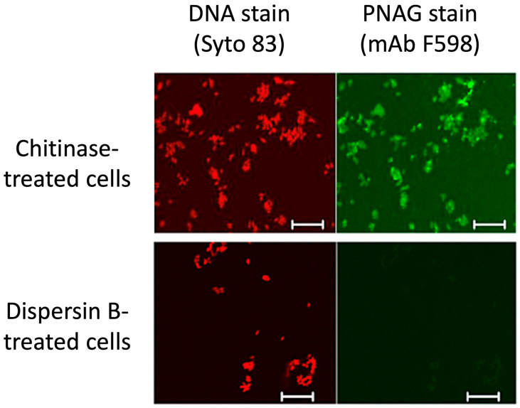
Figure 5.
Confocal microscopic analysis of PNAG expression by Y. pestis strain KIM6+ grown at 28 °C overnight on Congo red agar. After treatment of bacterial cells with either chitinase (top panels) or dispersin B (bottom panels), cells were stained with Syto 83 to visualize DNA (red) and Alexa Fluor 488-conjugated mAb F598 to detect PNAG (green). Bars = 10 µm. Figure from Yoong et al. [40].

