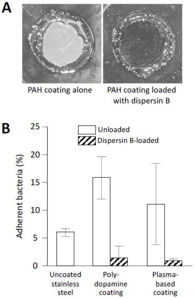
Figure 6.
Abiotic surfaces coated with dispersin B resist S. epidermidis biofilm formation and surface attachment: (A) Biofilm formation by S. epidermidis strain NJ9709 on glass slides containing an ultrathin layered poly(allylamine hydrochloride) (PAH) hydrogel coating (left panel) or a PAH coating loaded with dispersin B (right panel). Bacteria were cultured inside plastic cloning cylinders (5 mm internal diameter) that were attached to the slide with high-vacuum grease. After 12 h, the biofilms were rinsed, the cloning cylinders were removed, and the slides were photographed. The rings correspond to the footprints of the cloning cylinders. The biofilm appeared as a white film on the unloaded PAH layer, which was absent on the dispersin-B-loaded PAH layer. (B) Attachment of S. epidermidis strain ATCC35984 to uncoated stainless steel disks, or to disks coated with polydopamine- or plasma-based coatings with or without grafted dispersin B. Source: (A) [65]; (B) redrawn from [66].

