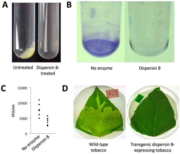
Figure 7.
Effects of dispersin B on plant-associated bacteria: (A) Xanthomonas citri subsp. citri strain 306 forms aggregates when cultured in broth (left panel). These aggregates were rapidly dissolved upon dispersin B treatment (right panel). (B) Biofilm formation by Ralstonia solanacearum strain Molk2 in polystyrene microtiter plates in the absence or presence of 20 µg/mL dispersin B. Biofilms were stained with crystal violet. (C) Binding of Pseudomonas fluorescens strain WCS365 to tomato roots in the absence or presence of 20 µg/mL dispersin B. Bacteria were mixed with 6-day-old tomato roots for 90 min. The roots were then crushed, mixed by vortex agitation, diluted, and plated on agar for CFU enumeration. Each data point represents one individual root. (D) Tobacco leaves infected with Pectobacterium carotovorum subsp. carotovorum strain ATCC 15713. Leaves were photographed 24 h after inoculation: (left) wild-type tobacco leaf; (right) leaf from a transgenic tobacco plant expressing dispersin B. Source: (A–C) J.B. Kaplan, unpublished data; (D) Ragunath et al. [104], N. Ramasubbu, unpublished data.

