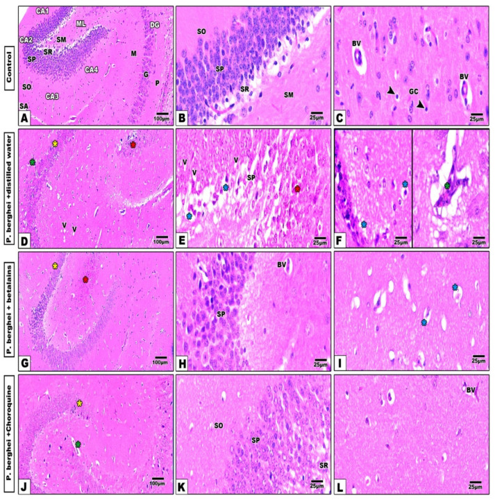
Figure 4.
Photomicrographs of hippocampus histological sections of control mice (Panel (A–C)) shows a well-organized regions architecture of the hippocampus. In infected mice treated with distilled water (Panel (D–F)), the hippocampus cell layers show several deformities especially in the proper (yellow asterisk) and dentate gyrus zones (red asterisk) with prominent vacuolation (V) and pyknosis (blue asterisks) in their cells and dilated capillaries (green asterisk). Treatment of infected mice with betalains (Panel (G–I)) or chloroquine (Panel (J–L)) display remarkable improvement in the histological architecture of hippocampus compartments in spite of little deformities still present especially in the cornu ammonis and dentate gyrus zones (H&E stain). Abbreviations: Blood vessel (BV), Cornu ammonis (CA), dentate gyrus (DG), molecular layer (ML), stratum alveolus (SA), stratum oriens (SO), stratum pyramidale (SP), stratum radiatum (SR), stratum molecular (SM), polymorph layer (P), granular layer (G), molecular layer (M), glial cells (GC) (arrow head).

