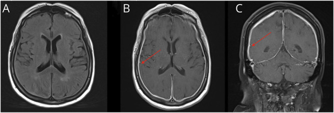
Figure 1. Brain MRI From November 2020.
This figure highlights our patient's brain MRI in November 2020. (A) A T2/FLAIR axial sequence of our patient, which notes no significant abnormality. However, on T1 postcontrast sequences, both axial cut (B) and coronal cut (C) note diffuse and smooth pachymeningeal thickening and enhancement (red arrows).

