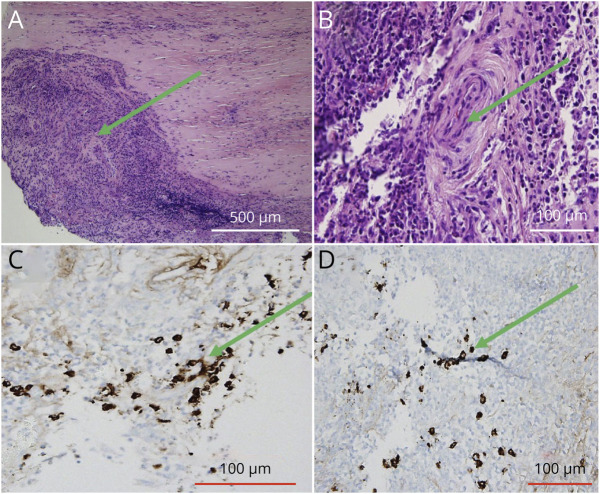
Figure 2. Dural Biopsy From November 2020.
This figure showcases our patient's dural biopsy. Her hematoxylin and eosin stains (A and B) are indicative of a substantial number of plasma cells and inflammatory infiltrates, with multinucleated giant cell formation (green arrows). There is no overwhelming disruption of blood vessels. The IgG4 stains (C and D) identify plasma cells that produce IgG4, but this histological slide is not suggestive of IgG4-RD.

