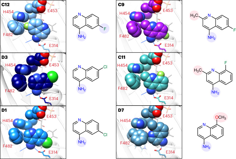
Fig. 6. Aminoquinoline active site orientations support TR-SAXS SAR.
Crystallographic complexes of CX1 and CX2 fragments with AIF(W196A) (C12, D3 and D1) or wild-type AIF (C9, C11 and D7) reveal stabilizing active site contacts and sources of steric interference that correlate with AIF dimerization rates. Spheres represent atomic van der Waals radii. Noncarbon atoms are colored dark blue (nitrogen), red (oxygen), light green (fluorine) and neon green (chlorine). Hydrogen bonds between the active site and primary amine are shown with blue lines. Substituents enhancing or hindering contact with the AIF active site are highlighted in blue and red, respectively, on the associated two-dimensional chemical diagrams. The images are ordered based on kVR ranking (see Fig. 3).

