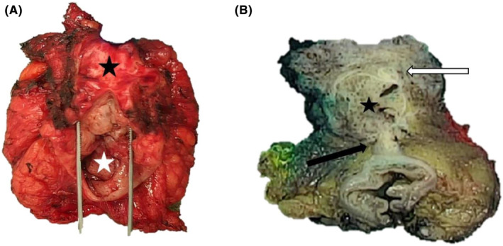
FIGURE 2.

Macroscopic photographs of the posterior pelvectomy with endometriosis lesions before and after formalin fixation. Gross findings of the resected specimen at first examination before fixation, with the vaginal wall (black star) attached to the rectum (white star) (A). Post‐fixation transversal section revealed a firm and gray‐tan, round cystic mass (star), located between and protruding through the vaginal chorion (white arrow) and the anterior rectal wall (black arrow) (B).
