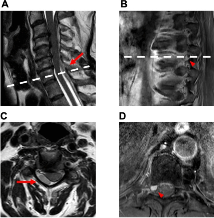
Fig. 6.
Case 4: 66-year-old man with spontaneous epidural hematoma. A 66-year-old male presented to the outpatient clinic for clinical and imaging follow-up. One month earlier, he had consulted the emergency department, experiencing pain between the shoulder blades. MRI revealed a spontaneous epidural hematoma from C2 to T3 (A + C), which was treated conservatively. At follow-up, the patient reported a complete resolution of his pain. A subsequent MRI showed a contrast-enhancing lesion at the T9 level on the right side (B + D). Upon discussion of the case in our neuro-oncology tumor conference, a resection of the lesion was recommended due to suspicion of a tumor. A preoperative DSA was not performed. The patient had no neurological deficits, and laboratory results were normal. Elective microsurgical excision of the extradural lesion was performed without any postoperative complications. The patient exhibited no sensorimotor symptoms. Histopathological examination confirmed the lesion as a hemangioma. The patient was discharged on the second postoperative day

