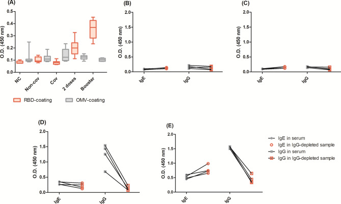
Fig. 6.
Control tests: (A) sera background comparing the optical density (OD) of IgE-detection incubating sera at 1:5 dilution in microplates coated with SARS-CoV-2 RBD or N. meningitidis OMVs. Each group (negative control, Non-cov, Cov, 2 doses and booster) had an n = 8. For another control assay, we depleted IgG from the samples, to test if, once depleted, the IgE optical signal (using anti-IgE-ε chain) would be maintained after depletion whereas the IgG optical signal (using anti-IgG-Fc) would decrease after depletion. Four samples were used: (B) collected before the pandemic as negative controls, (C) collected before COVID-19 vaccines were available. In these cases, there were no specific antibodies in the samples, and the O.D.s, before and after depletion, were almost the same. Four samples (D) collected after two vaccine doses, and (E) collected after the booster dose. In these cases, there was both IgG and IgE in the samples, so the IgE O.D. was either the same or even enhanced, probably due lack of IgG competition for RBD, while IgG O.D. decrease, proving that the depletion worked.

