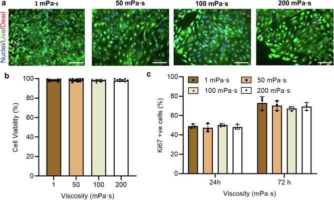
Fig. 2. Effect of culture media viscosity on FTEC viability and proliferation.
a Representative live/dead (green/red) fluorescence staining images of cells at the 72-h time point and the b percentage of live cells (cell viability). Cell nuclei were stained using blue-fluorescent Hoechst 33342 (n = 9 images from three biological replicates per each condition). Scale bar, 50 µm. c The percentage of Ki-67 positive cells at the 24-h and 72-h time points. Experiments were carried out on n = 3 from three biological replicates and ≥ 200 cells for each condition were analysed. All data are represented as mean ± s.d. and analysed using one-way ANOVA with Tukey post hoc testing. Source data are provided as a Source Data File.

