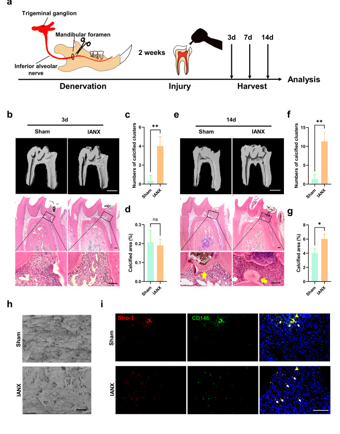
Fig. 1.
Denervation of sensory nerves leads to poor pulp repair and ectopic mineralization. a Schematic illustrating the denervation and pulp injury strategy (n = 4). b MicroCT and HE staining of the Sham and IANX groups at 3 d. MicroCT scale bar = 1 mm; HE scale bar = 100 μm. c Quantification of the calcified clusters number of MicroCT in b (**P < 0.01, n = 3). d The percentage of the calcified area of the total pulp area (%) of MicroCT in b (ns indicates P ≥ 0.05, n = 3). e MicroCT and HE staining of the Sham and IANX groups at 14 d. Yellow arrows indicate mineralized tissue. MicroCT scale bar = 1 mm; HE scale bar = 100 μm. f Quantification of the calcified clusters number of MicroCT in e (**P < 0.01, n = 3). g The percentage of the calcified area of the total pulp area (%) of MicroCT in e (ns indicates P ≥ 0.05, n = 3). h Scanning electron microscopy of restorative dentin bridges in the Sham group and mineralized masses in the IANX group at 14 days after pulp injury as indicated by yellow arrows in c. Scale bar = 20 μm. i Immunofluorescence staining of Stro-1(red) and CD146 (green) in the pulp close to the injury of the Sham and IANX groups at 3 d. Images are at a similar magnification with the lower panel of b. White arrows indicate co-staining. Yellow arrows indicate dental pulp injury sites. Scale bar = 100 μm

