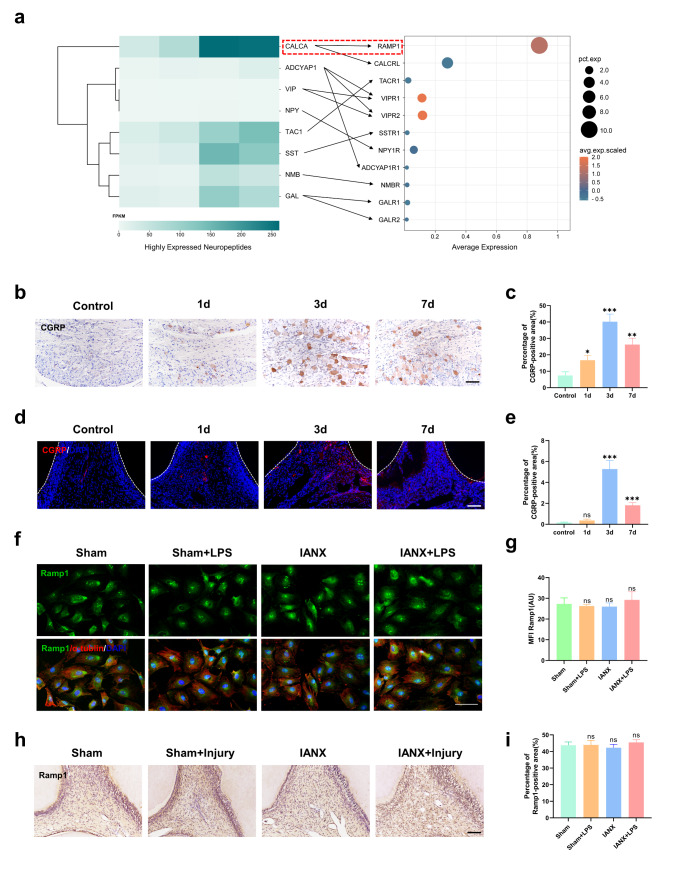
Fig. 3.
Sensory nerves act directly on DPSCs through the CGRP-Ramp1 axis. a Left: Heatmap of the top-expressed neuropeptides in the trigeminal ganglion; Right: Dotplot of respective cognate receptor expression (denoted by arrows) in DPSCs at steady state (circle diameter reflects the percentage of cells expressing, circle color reflects relative expression abundance). b Immunohistochemical staining of CGRP in the trigeminal ganglion after pulp injury. Scale bar = 100 μm. c Statistical analysis of CGRP-positive area in the trigeminal ganglion (*P < 0.05, **P < 0.01, ***P < 0.001, n = 3). d Immunofluorescence imaging of CGRP in dental pulp after injury. Scale bar = 100 μm. e Statistical analysis of CGRP-positive area in the dental pulp (ns indicates P ≥ 0.05, ***P < 0.001, n = 3). f Immunofluorescence staining of Ramp1 (green) and α-tubulin (red) in the DPSCs of Sham and IANX groups. Scale bar = 100 μm. g Statistical analysis of MFI (Mean Fluorescence Intensity) in d. (ns indicates P ≥ 0.05, n = 15). h Immunohistochemical staining of Ramp1 in the dental pulp after injury. Scale bar = 100 μm. i Statistical analysis of Ramp1-positive area in the dental pulp (ns indicates P ≥ 0.05, n = 3)

