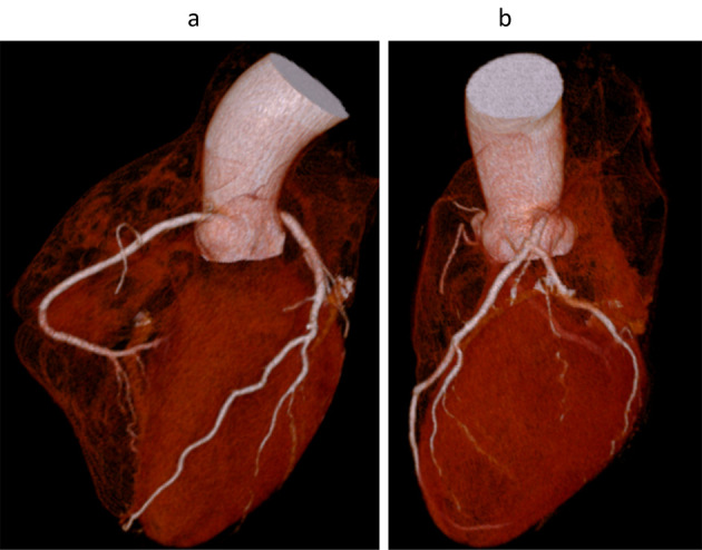
Figure 2.

Three-dimensional volume rendering images acquired with 64-slice coronary computed tomography angiography of the right (a) and left (b) coronary arteries of the patient. There was no obvious coronary artery stenosis in either the right or the left coronary arteries.
