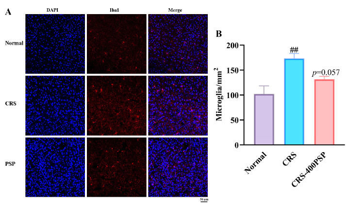
Figure 4.
PSP attenuated microglial activation in the prefrontal cortex of CRS-induced mice (n = 5). (A) Immunofluorescence staining of microglia: representative images showing immunofluorescence staining of microglia in the prefrontal cortex. Blue indicates DAPI-stained nuclei, and red indicates Iba-1-stained microglia. Scale bar: 50 μm. (B) Quantification of microglial number per square millimeter. ## p < 0.01 vs. normal group.

