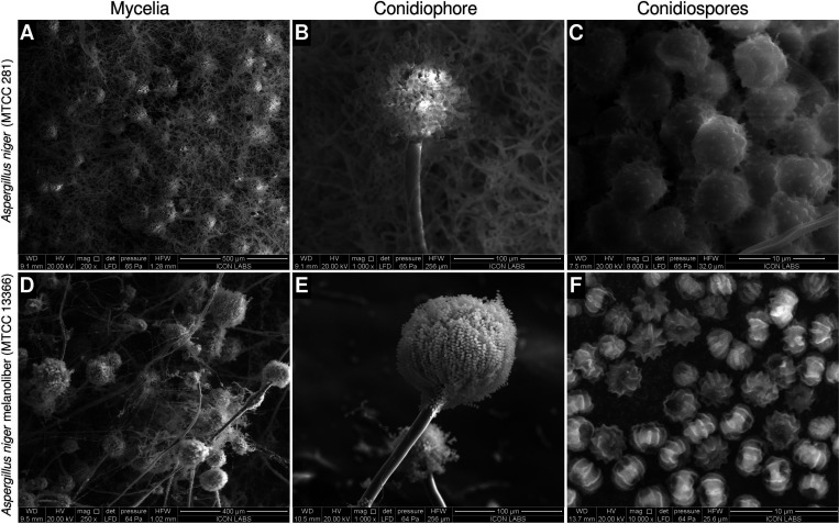
Figure 1.
Morphology of Aspergillus niger strains used in this study. (A–C) The A. niger type strain (MTCC 281). (A) Dense hyphal mass possessing abundant conidiophores. (B) Conodiophore possessing relatively fewer conidiospores (C) Large conidiospores with echinulations evenly distributed across their surface. Polar regions are not discernible. (D–F) A. niger melanoliber (MTCC 13366). (D) Sparse hyphal mass possessing fewer large conidiophores. (E) Conodiophore possessing a large number of small conidospores. (F) Small conidiospores possessing deep, longitudinal striations originating from discernible polar regions.

